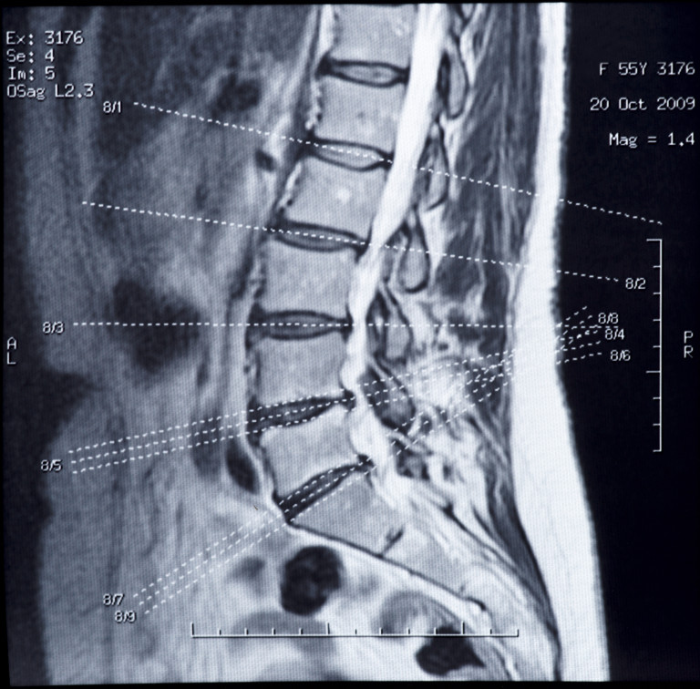
 When do doctors recommend an MRI for scoliosis? Typically MRIs are diagnosed for patients diagnosed in the juvenile phase (<ten-years-old) or for adolescents experiencing atypical characteristics of idiopathic scoliosis.
More specifically, an MRI for scoliosis may be recommended when a patient has numbness, weakness, pain, or an asymmetric loss of reflexes. An MRI can help rule tethered cord syndrome in or out, and congenital malformations such as a syrinx and/or Chiari malformations.
A tethered cord is a neurological disorder that occurs when the spinal cord is abnormally attached to tissues around the spine. This results in limited spinal cord movement within the spinal column, which can sometimes cause nerve damage. Syrinxes are fluid-filled cavities that can form in the spinal cord or brain stem. Chiari malformations are structural defects in the cerebellum – the part of the brain that controls balance.
An MRI for scoliosis may also be recommended when a child is diagnosed with a severe curve of sudden onset or when scoliosis progresses rapidly. An MRI is usually ordered for an atypical curve pattern such as a thoracic levoscoliosis (i.e., a left thoracic curve). Thoracic levoscoliosis is uncommon and estimated at only 10% of all scoliosis curves.
In addition to the aforementioned circumstances, an MRI is usually always ordered when a scoliosis is diagnosed in childhood between 4 and 10 years old–which is about 10-15% of cases diagnosed.
An MRI is usually ordered before spinal fusion surgery. A recent study found that 27% of patients undergoing surgery did exhibit neuro-axial anomalies that were previously undetected clinically and radiographically (1). Your doctor will order an MRI if he/she feels it is necessary, but if you have concerns, seek out a second opinion.
1 – Rajasekaran S, Kamath V, Kiran R, Shetty AP. Intraspinal anomalies in scoliosis: An MRI analysis of 177 consecutive scoliosis patients. Indian Journal of Orthopaedics. 2010;44(1):57-63. doi:10.4103/0019-5413.58607

