Here at Scoliosis 3DC® (3DC stands for three-dimensional correction), successful conservative management of scoliosis includes exercises for the sagittal plane. From the onset, our program has integrated sagittal plane exercises on the basis that sagittal plane instability may be a contributing cause of scoliosis. Sagittal plane exercises are used so patients learn to work toward halted curve progression, or optimally, to achieve some degree of scoliosis correction. To this end, some recent research out of Children’s Hospital of Philadelphia caught our attention.
A study by Saba Pasha, entitled 3D Deformation Patterns of S Shaped Elastic Rods as a Pathogenesis Model for Spinal Deformity in Adolescent Idiopathic Scoliosis (1), now supports and expands upon previous publications in regard to the sagittal plane, scoliosis, and perhaps one potential explanation regarding the cause of scoliosis.
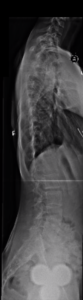
In an attempt to determine a cause of scoliosis, the author used the sagittal plane x-rays of 126 adolescent females with right thoracic scoliosis (the most common type) and compared them to the sagittal plane x-rays of 3 non-scoliotics. “Computer simulations were then used to investigate how elastic rods, modeling children’s spines, change shape in response to mechanical loading.” Her study concludes that “the shape of a person’s sagittal profile can be a leading cause of scoliosis” (1). This confirms a premise we’ve taken into account when treating scoliosis, for years!
The author explains, “The findings of this study reduced the question of why scoliosis happens to why variation in the sagittal spinal profile occurs in the pediatric population.” The study also states that “the current analysis can be used as a risk assessment tool to predict the risk of spinal deformity development in the pediatric population. This can open a new line of research that aims to prevent the spinal deformity development by determining the spinal loading patterns (the sagittal alignment and flexibility of the immature spine) at a higher risk of 3D deformation in pre-scoliotic pediatric population” (1).
She also asserts, “Conservative methods to protect the spine during this phase can be explored,” and contends that continued research is needed to develop “strategies to prevent a condition for which no preventive measures now exist” (1). Yet, they do here at Scoliosis 3DC as we address and have always addressed the sagittal plane of scoliosis as part of our comprehensive program.
One component of our program shows patients how to integrate proactive strategies to positively influence the sagittal plane. The overall goal is to create spinal stability where instability exists in a scoliotic (Cobb angle of 10º or more) or pre-scoliotic spine (Cobb angle under 10º). The purpose of our approach is to help growing children avoid spinal curve progression or ideally, to improve children’s spines as they grow and develop.
This is accomplished via easy sagittal plane exercises, a simple yet important part of our scoliosis program. When these exercises were studied in 2006(1). It was concluded that sagittal plane correction exercises are, “a useful ‘add-on’ to scoliosis rehabilitation with regard to the lateral deviation of the scoliotic trunk in the short-term.” It was also noted that “correction forces applied in the sagittal plane are also able to correct the scoliotic deformity in the coronal plane”(2). In addition, active self-correction techniques(3), when applied, enable the patient to modify their regular postural stances to reduce asymmetric spinal loading – a major contributor to curve progression (4).
Pasha goes on to suggest that for those at risk, perhaps “wearing a brace at a younger age may prevent scoliosis from developing” (1). Again, this is and has been our philosophy here at Scoliosis 3DC® for years. It’s what many parents who prefer proactive treatment for scoliosis opt for rather than abiding by the watch-and-wait philosophy that is so common in traditional medical circles! This is especially true for young patients with a high risk of progression, or a family history of scoliosis.
In 2008, van Loon et al. provided evidence of influencing the sagittal plane with regard to scoliosis bracing. Their conclusion: “Scoliosis deformities are significantly reduced in the supine position by a lordotic fulcrum force on the thoracolumbar junction and that these findings may have an impact on bracing techniques”(5). In a follow-up study, it was concluded that “significant reduction of scoliosis and kyphotic curves is possible during growth. By stepwise restoration of thoracolumbar lordosis and preventing overload in compression during sitting creates improved condition”(6).
Sagittal plane flattening is something we see often and view as a possible cause of scoliosis as well as a potential indicator of future progression potential. Dr. Marc discusses this in his What is Scoliosis? YouTube video. The Scoliosis 3DC® spinal management protocols for the sagittal plane teach patients at risk with pre-scoliosis (curves under 10º), as well as those with mild, moderate, and severe scoliosis, how to incorporate simple exercises into their lives to stabilize the spine. When it comes to treating scoliosis conservatively, when the right protocols are implemented, sooner rather than later, they have the potential to be more impactful. It’s one reason our tagline is Scoliosis3DC®, Not wait and see!
1) Pasha, S. 3D Deformation Patterns of S Shaped Elastic Rods as a Pathogenesis Model for Spinal Deformity in Adolescent Idiopathic Scoliosis. Sci Rep 9, 16485 (2019).
2) Weiss HR, Klein R. Improving excellence in scoliosis rehabilitation: a controlled study of matched pairs. Pediatr Rehabil 9: 3. 190-200 Jul/Sep 2006.
3) Monticone M, Ambrosini E, Cazzaniga D, Rocca B, Ferrante S. Active self-correction and task-oriented exercises reduce spinal deformity and improve quality of life in subjects with mild adolescent idiopathic scoliosis. Results of a randomised controlled trial. Eur Spine J 2014 Feb 28.
4) Stokes IA, Burwell RG, Dangerfield PH; IBSE. Biomechanical spinal growth modulation and progressive adolescent scoliosis–a test of the ‘vicious cycle’ pathogenetic hypothesis: summary of an electronic focus group debate of the IBSE. Scoliosis. 2006;1:16. Published 2006 Oct 18.
5) van Loon P.J., Kühbauch B.A., Thunnissen F.B. Forced lordosis on the thoracolumbar junction can correct coronal plane deformity in adolescents with double major curve pattern idiopathic scoliosis. Spine. 2008;33(7):797–801.
6) van Loon, P.J., Roukens, M. & Wever, D. Brace treatment with progressive lordotic forces at the thoracolumbar junction in adolescent scoliosis and hyperkyphosis. Scoliosis 4, O47 (2009).